Structural study of lipid droplet using synchrotron label-free multimodal imaging
Résumé
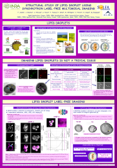
Imaging intracellular compartments, their dynamics and interactions in living cells, remains challenging. Using various lipid droplet (LD) protein markers, we revealed inter LD heterogeneity at single cell level, due to LD specific geolocalisation and enzymatic equipment. Even so, using tagged proteins or vital probes could modify the morphology and the smooth running of the organelles. Methods based on intrinsic fluorescence of molecules upon excitation by deep ultra violet (DUV) illumination are thus emerging for living cell imaging. We used DUV from synchrotron radiation to perform autofluorescence and transmittance imaging on single living yeast. The contrasted signals inside the cells revealed chemical heterogeneity at the subcellular level. Microscopy showed organelles with low auto-fluorescence after DUV illumination. We distinguished two populations, with high or low transmittance. The first population corresponded to vacuoles and the second to LDs. LDs appeared as heterogeneous well-organized structures with a low transmittance zone on the surface and a high transmittance core. We propose that the low transmittance ring and the high transmittance core correspond to ergosterol and triacylglycerol-containing structures, respectively. The conclusions we drawn using DUV imaging were confirmed by experiments performed using soft X ray imaging on cryofixed cells. Synchrotron label free imaging paves the way for efficient structural and dynamic studies of LDs and other organelles.
Fichier principal
 2018_Jamme_Poster_PhysBioSys_1.pdf (1.19 Mo)
Télécharger le fichier
2018_Jamme_Abstract_PhysBio_2.pdf (369.78 Ko)
Télécharger le fichier
2018_Jamme_Poster_PhysBioSys_1.pdf (1.19 Mo)
Télécharger le fichier
2018_Jamme_Abstract_PhysBio_2.pdf (369.78 Ko)
Télécharger le fichier
| Origine | Fichiers éditeurs autorisés sur une archive ouverte |
|---|
| Origine | Fichiers produits par l'(les) auteur(s) |
|---|
Loading...