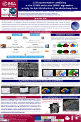
2.5D Representations Combining in vivo 3D MRI and ex vivo 2D MSI Approaches to Study the Lipid Distribution in the Whole Sheep Brain
Résumé
Mass Spectrometry Imaging (MSI) provides easily high spatially resolved masses allowing characterization of endogenous lipids. These latter constitute about 70% of the composition of the white matter of the brain which can be implicated in developmental and/or cognitive troubles. In order to examine the molecular distribution of lipids in whole sheep brain, and especially in white/grey matter, we combined in vivo and ex vivo images, obtained in the same animals, using Magnetic Resonance Imaging (MRI) and MSI, respectively. In order to view the topology of the molecular species within the organ, we propose the construction of a 2.5 D representation where a single section imaged with 2D MSI is localized within the tissue volume obtained by 3D MRI.
3D T1-weighted MPRAGE images were acquired on two anesthetized sheep with a 3 Tesla MRI (Siemens, Verio ®). The parameters of acquisition for the MPRAGE were: TR 2500ms, TE 3.2ms, FA 12, NEX 1, matrix 384×384, FOV 192mm, 288 slices with a thickness of 0.5mm. In order to improve data quality, the 3D MRI volumes have been pre-processed using in-house algorithms using volume fitting and Markov random field methods. T1 3D planes corresponding to MSI planes were reconstructed using Osirix imaging software.
Brains were collected after sacrifice and frozen at -80°C. Frontal and sagittal 14 µm brain sections were performed with a cryostat adapted to large sections (CM3050 S, Leica) and mounted onto conductive ITO-coated slides. The spray of α-cyano-4-hydroxycinnamic acid matrix was performed using an Image Prep device (Bruker). Spectra were acquired using an UltrafleXtrem MALDI-TOF instrument (Bruker) in the 200–1200 m/z range with a spatial resolution set at 125 µm. Raw spectra were analyzed with SCiLS Lab software to generate 2D ion density maps and segmentation maps (data partitioning).
The tissue sections analyzed by MSI were stained with cresyl violet to manually delimitate neuronal nuclei and areas. This histological map was used to delineate the MRI and MSI 2D views and overlay them regardless the same brain areas used as fiducials. After, a 2.5 D representation was proposed to visualize the lipid distribution within the entire organ.
In conclusion, in this study, frontal and sagittal whole sheep brain sections analyzed by MSI showed a clear difference in lipid distribution between different compartments of brain tissues, especially between grey and white matter, until the cerebral envelopment presenting circumvolution. Furthermore, the alignment of 2D MALDI-imaging with T1-weighted images showed that MSI can provide finer details on the structural connectivity of myelinated fiber tracts. Here, the 2.5 D representation combining MRI and MSI was presented as an alternative approach to 3D anatomical and molecular atlas providing a perfect topology of the molecular species within an organ. For the moment, 3D MSI of whole sheep brain is a challenge, while the 2.5 D construction demonstrated to be a capable tool for exploring molecular distributions throughout sample volumes.
Nowadays, the reported results may serve as a starting point for further experiments associating MSI and dynamic and functional MRI, especially for the characterization of brain.
Fichier principal
 Poster 2.5 D representation whole brain_lipidomic_final.pdf (3.69 Mo)
Télécharger le fichier
Poster 2.5 D representation whole brain_lipidomic_final.pdf (3.69 Mo)
Télécharger le fichier
Origine : Fichiers produits par l'(les) auteur(s)