TUMOR MICROENVIRONMENT IMAGING: BENEFITS OF MULTIMODALITY TO STUDY CHONDROSARCOMA
Résumé
INTRODUCTION: Chondrosarcoma (CHS) is a malignant cartilaginous tumor representing the most common primary bone cancer in adults.[1] Due to its dense chondrogenic extracellular matrix and hypoxic environment, CHS is highly resistant to conventional chemotherapy and radiation.[2] Development of multimodal imaging to characterize and map in vivo CHS microenvironment is fundamental for specific diagnosis and personalized therapy. In this work, we proposed to combine the resolution of chemical exchange saturation transfer (CEST) MRI with nuclear imaging sensitivity to improve CHS microenvironment understanding.[3]
METHODS: Swarm rat CHSs were implanted subcutaneously in NMRI nude mice (n=10). When tumors were measurable (12-16 days post-transplant), mice were imaged by CEST MRI.[4] Proteoglycans, the main component of chondrogenic extracellular matrix, were quantified by GAG CEST contrast. Guanidyl-and APT CEST contrasts were combined to characterize acidic pH, as hypoxia reflect. These two features, proteoglycans and hypoxia, were assessed in parallel by nuclear imaging with 99m Tc-NTP 15-5 SPECT imaging [5] and 18F-FMISO PET imaging [6], respectively. Data were also completed by ex vivo analyses of proteoglycans (Alcian blue stain and biochemical assay with dimethylmethylene blue) and hypoxia (pimonidazole immunofluorescence).
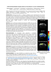
RESULTS/DISCUSSION: For extracellular matrix assessment, a proteoglycan concentration-related map was established with GAG CEST contrast showing more intense areas in the tumor than in the muscle (AUC =27.29 ± 8.38 vs 2.15 ± 1.88, respectively; P < 0.0001) (Fig 1). Similarly, a significant (P < 0.0001) radiotracer uptake was observed by nuclear imaging with 99mTc-NTP 15-5 within tumoral tissue (179.38 ± 38.39 kBq/mL) as opposed to muscle (35.07 ± 5.17 kBq/mL). These results were supported by biochemical assay (6.33 ± 1.60 μg/mg vs 0.41 ± 0.09 μg/mg, respectively; P < 0.0001). Considering hypoxia, a small discrepancy was observed between CHS and muscle with 18F-FMISO PET imaging. Interestingly, with Guanidyl and APT contrast, the weakly hypoxic status of the CHS was explained by a heterogeneous acidic pH distribution within the tumoral tissue (Fig 1). Moreover, immunofluorescence with pimonidazole evidenced a weak staining within tumor mainly localized in the center of CHS lobules.
CONCLUSION: The results from CEST MRI, nuclear imaging and ex vivo analyses were in agreement and highlighted a rich proteoglycan extracellular matrix and a heterogeneous hypoxic tumoral microenvironment for Swarm rat CHS xenograft in mice. This study emphasizes the role of multimodal imaging to characterize tumor phenotypes resistant to treatments and allows a better understanding of the relationship between tumor cells and their environment.
Domaines
Cancer
Fichier principal
 7_Autissier.pdf (575.58 Ko)
Télécharger le fichier
JGS 2021 - Poster.pdf (1.71 Mo)
Télécharger le fichier
P7_Autissier.pdf (915.72 Ko)
Télécharger le fichier
7_Autissier.pdf (575.58 Ko)
Télécharger le fichier
JGS 2021 - Poster.pdf (1.71 Mo)
Télécharger le fichier
P7_Autissier.pdf (915.72 Ko)
Télécharger le fichier
| Origine | Fichiers produits par l'(les) auteur(s) |
|---|